Advanced materials characterization enhanced by cutting-edge techniques
These advanced instruments will not only support MEL’s research but will also be accessible to other REFRESH living laboratories, various research groups at VSB-TUO, international collaborators, and commercial partners.
“The development of novel materials demands comprehensive chemical, structural, morphological, and physico-chemical characterization to understand the connections between the synthesis conditions and the resulting chemical, structural and application-relevant properties,” explained the MEL manager Ondřej Malina.
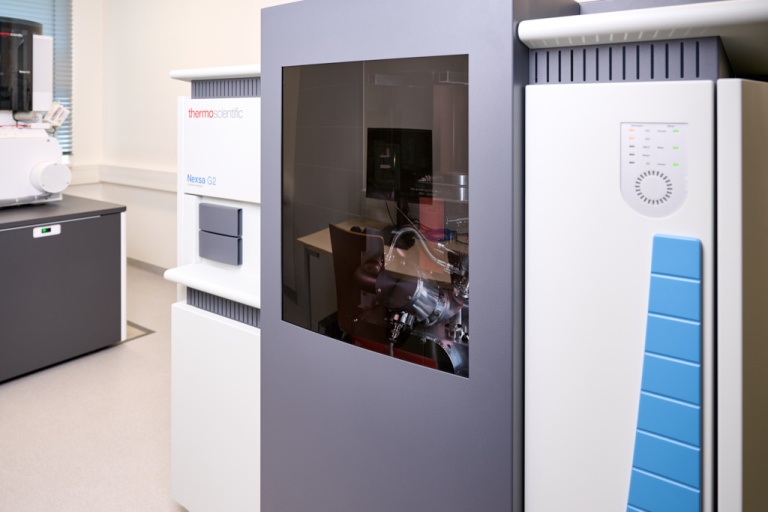
X-ray photoelectron spectroscopy (XPS) is a powerful technique for chemical and structural analysis of solid surfaces, including thin films, coatings and powdered materials. This advanced system is designed for highly precise quantitative and qualitative chemical analysis. It also enables differentiation of oxidation states in metals and identification of organic functional groups within material structures. Moreover, scientists can utilize depth profiling to gain unique insights into compositional and structural variations both at the surface and within the bulk of materials.
“The new XPS instrument in our laboratory offers a truly unique integration with a high-resolution scanning electron microscope, allowing for the simultaneous analysis of chemical and structural properties within precisely defined sample regions. Beyond chemistry and structure, this system also provides valuable data on particle size and morphology in a single analytical session. This combination of techniques is unparalleled on a European scale, and we anticipate significant interest in its capabilities—not only within VSB-TUO but also from domestic and international academic institutions and commercial partners,” added the MEL Head Radek Zbořil.
The high-resolution scanning electron microscope represents a state-of-the-art solution for detailed size, morphological and structural analysis of advanced materials. At MEL, experts employ this instrument to characterize low-dimensional materials, including graphene derivatives, metal-organic frameworks, metal oxide nanotubes and carbon dots, which have promising applications in green energy, catalysis and medicine. Additionally, the system enables combined scanning and transmission electron microscopy (STEM) imaging, facilitating precise chemical mapping of selected sample regions. Beyond research, this sophisticated technique is indispensable to numerous commercial partners seeking high-resolution material characterization services.
Text: Martina Šaradínová
Photo credit: Petr Havlíček