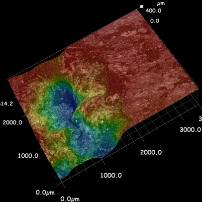
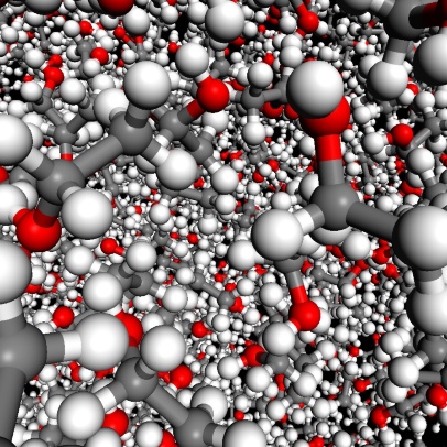
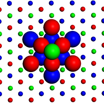
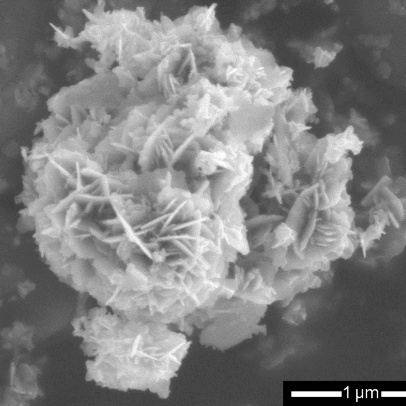
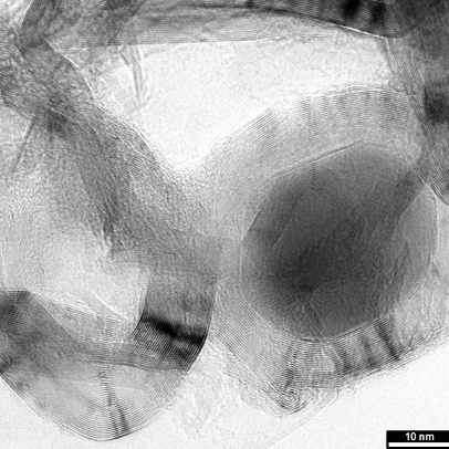
In our laboratory we focus on syntheses of nanocomposite materials, the study of their structure and properties. In syntheses, we place emphasis not only on reproducibility, but also on ecological safety and economic efficiency. We characterize the structure of nanomaterials with a wide range of instrumental methods, but especially with X-ray diffraction analysis and Raman and infrared spectroscopy. To study the microstructure, we use digital microscope able to create a 3D views, to measure optical properties, we use UV-VIS and diffuse reflectance spectrophotometry. Photocatalytic activity and electrical conductivity are measured using established methods. We supplement the experimental data with the results of computer simulations based on molecular mechanics and dynamics, including simulated X-ray diffraction. In our research, we cooperate with a number of laboratories not only at VŠB-TUO.
Research focus
- Nanocomposites based on clay minerals modified with photoactive nanoparticles (TiO2, ZnO, ZnS).
- Nanocomposites based on clay minerals intercalated with conductive polymers (polyaniline, polypyrrole).
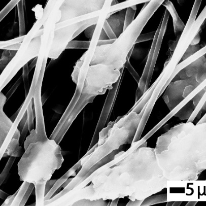
- Surface-modified nanofibers.
- Long-term monitoring of changes in the properties of nanomaterials.
- Impact of nanomaterials on the environment and their interaction with organisms.
Offered services
- X-ray diffraction analysis
- Fourier transform infrared spectroscopy
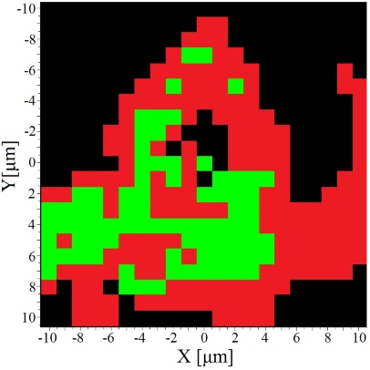
- Raman spectroscopy
- UV-VIS spectroscopy and DRS (diffusion reflectance) spectrophotometry
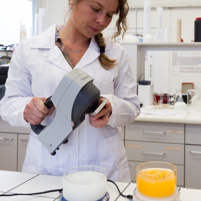
- Optical microscopy
- Biodeterioration - determination of the resistance of surfaces to overgrowth with biofilm
- Determination of acute toxicity using green algae
- Determination of photocatalytic activity using established method
Contact
doc. Ing. Jonáš Tokarský, Ph.D.
e-mail:
phone: +420 597 321 606; +420 597 321 519
